RABEA® — Cervical fusion cage
Anterior cervical fusion cage (ACDF) with an open rectangular design — an established SIGNUS standard, available in PEEK-OPTIMA® or titanium, for the C3–TH1 segment.
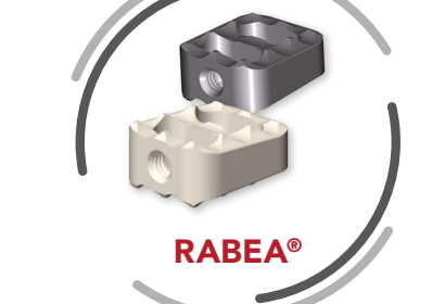
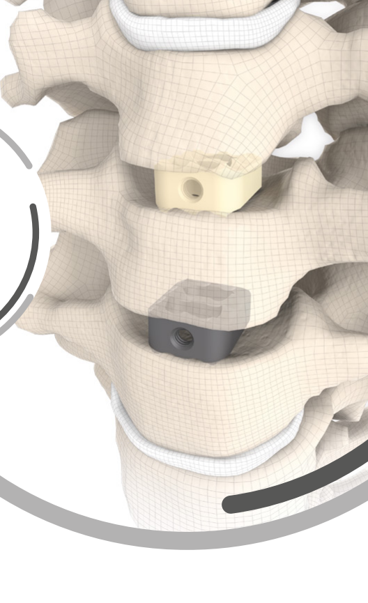
RABEA® is an anterior cervical intervertebral fusion cage developed by SIGNUS for the specific demands of anterior cervical fusion (C3–TH1). It is implanted using the established Smith–Robinson technique. With CE marking since 1996, RABEA® remains, after more than 25 years, a standard in anterior cervical fusion.
The open rectangular design allows the cage to be filled with natural bone graft or with a synthetic substitute (for example KAINOS® Inject), promoting osseointegration. The serrated teeth characteristic of SIGNUS provide firm anchorage in bone, with high primary stability and a reduced risk of implant migration. The wide selection of implants offers intraoperative flexibility and restoration of the intervertebral space.
RABEA® is available in high-performance PEEK-OPTIMA® or in solid titanium (Ti-6Al-4V). The radiolucent PEEK variant has titanium alloy radiographic markers (anterior and posterior) for intra- and postoperative verification and allows artifact-free MRI imaging, for optimal monitoring. Cages with a 5° lordotic angle are available, as well as plane-parallel implants (0°). Implantation requires a single instrument. For additional anterior fixation, the TOSCA® or ASCOT® ventral plates can be used.
- Degenerative disorders of the cervical disc, at the C3–TH1 level.
- Disc herniation; mechanical instability; osteochondrosis.
- Spinal canal stenosis; spondylolisthesis.
- Pseudarthrosis or failed anterior spondylodesis (fusion).
Rectangular design
Compatible with the established Smith–Robinson technique, for a familiar surgical workflow.
Open design
Filled with natural bone graft or synthetic substitute (e.g. KAINOS® Inject) and promotes osseointegration.
SIGNUS serrated teeth
Firm anchorage in bone, with high primary stability and a reduced risk of implant migration.
Artifact-free MRI
In the PEEK variant — optimal postoperative monitoring; titanium radiographic markers for positioning.
Two materials
Radiolucent PEEK-OPTIMA® or solid titanium (Ti-6Al-4V), depending on clinical preference.
A single instrument
Implantation with a single inserter — simple, safe, and economical handling.
- Type
- Anterior cervical intervertebral fusion cage (ACDF), open, rectangular design
- Level
- Cervical spine C3–TH1; Smith–Robinson technique
- Material
- PEEK-OPTIMA® (radiolucent, with titanium markers Ti-6Al-4V) or solid titanium (Ti-6Al-4V)
- Footprints (width × length)
- 12×12 mm and 12×14 mm
- Heights
- 4–8 mm (depending on footprint and material; excluding tooth height)
- Lordosis
- 0° (plane-parallel) and 5°
- Teeth (serration)
- 1 mm on each face
- Filling
- Natural bone graft or synthetic substitute (e.g. KAINOS® Inject)
- Optional anterior fixation
- TOSCA® or ASCOT® ventral plates
- Instrumentation / supply
- A single inserter; single-use, sterile implants
Would you like information about RABEA® — Cervical fusion cage?
The Ronda Medical team provides details about availability, configurations, and technical support.
Contact the team